41 diagram of the lungs with labels
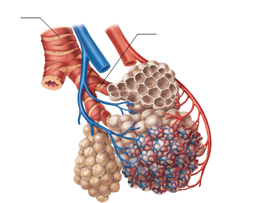
Label the lung diagram - Quizlet Start studying Label the lung diagram. Learn vocabulary, terms, and more with flashcards, games, and other study tools. Lungs (Human Anatomy): Picture, Function, Definition, Conditions - WebMD The lungs are a pair of spongy, air-filled organs located on either side of the chest (thorax). The trachea (windpipe) conducts inhaled air into the lungs through its tubular branches, called...
Label Lungs Diagram Printout - EnchantedLearning.com Read the definitions below, then label the lung anatomy diagram. bronchial tree - the system of airways within the lungs, which bring air from the trachea to the lung's tiny air sacs (alveoli). cardiac notch - the indentation in the left lung that provides room for the heart. diaphragm - a muscular membrane under the lungs.
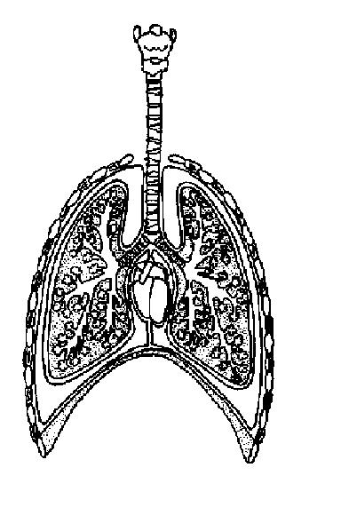
Diagram of the lungs with labels
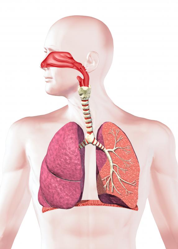
Label Lungs Diagram Printout - EnchantedLearning.com | Respiratory ... Diagram illustrating the paths of blood flow through the heart. Blood enters the heart through two large veins, the inferior and superior vena cava, emptying oxygen-poor blood from the body into the right atrium. Label Lungs Diagram Printout - EnchantedLearning.com Read the definitions below, then label the lung anatomy diagram. bronchial tree - the system of airways within the lungs, which bring air from the trachea to the lung's tiny air sacs (alveoli). cardiac notch - the indentation in the left lung that provides room for the heart. diaphragm - a muscular membrane under the lungs. Lungs: Definition, Location, Anatomy, Function, Diagram, Diseases Where are the Lungs Located. The lungs are located a little toward the posterior part of the human body, just below the collarbone, extending down to the diaphragm, the muscular partition that separates the chest and abdominal cavities.The left and right lungs are situated on the two sides of the body with the heart, another vital organ in the thoracic cavity, located a little in front of, and ...
Diagram of the lungs with labels. Diagram of Human Heart and Blood Circulation in It Four Chambers of the Heart and Blood Circulation. The shape of the human heart is like an upside-down pear, weighing between 7-15 ounces, and is little larger than the size of the fist. It is located between the lungs, in the middle of the chest, behind and slightly to the left of the breast bone. The heart, one of the most significant organs ... Labeled Diagram of the Human Lungs - Bodytomy Given below is a labeled diagram of the human lungs followed by a brief account of the different parts of the lungs and their functions. Each lung is enclosed inside a sac called pleura, which is a double-membrane structure formed by a smooth membrane called serous membrane. Label Diagram Of The Lungs - Solved Labeling Diagram Directions Label ... This fantastic lung labelling activity provides learners with a blank lung diagram for them to label themselves. Alveoli, trachea, bronchi, bronchioles, diaphragm, lung, mouth, and nasal passage,. Make sure you know the basics of lung cancer, including prevention, risk factors, symptoms and treatment options. Label lungs anatomy diagram printout. Human Lungs Diagram - Agaliprogram Given below is a labeled diagram of the human lungs followed by a brief account of the different parts of the lungs and their functions. Each lung reaches from the collarbone to the border between the chest and abdominal cavities. Source: en.wikipedia.org An illustration depicting cigarettes and a pair of lungs affected by smoking.
Lungs label - Teaching resources 3728 results for 'lungs label'. Lungs Labelled diagram. by Rbowerkail. KS4 PE. The Lungs Labelled diagram. by Fayeroberts. KS4 Y10 Biology. Lungs Diagram Labelled diagram. by Jon9. The Respiratory System (Label Diagram) - ScienceQuiz.net Match each pair by dragging from right to left. When complete click Check button. Labeling Diagrams Of The Heart - Isacork 1.Different Parts Of The Body ↓ 2.Major Veins ↓ 3.Right Atrium ↓ 4.Right Ventricle ↓ 5.Pulmonary Artery ↓ 6.Lungs The two upper chambers are called the left and the right atria, and the two lower chambers are known as the left and the right ventricles. The boxes are numbered to correlate with the labeled chambers on the cartoon diagram. Label the Lungs Diagram | Quizlet ... superior lobe of right lung ... middle lobe of right lung ... inferior lobe of right lung ... superior lobe of left lung ... left main (primary) bronchus ... lobar (secondary) bronchus ... segmental (tertiary) bronchus ... inferior lobe of left lung ... Sets found in the same folder Bi 233: Labeling the Larynx 21 terms SunshineGirl79 the cell
Lobes of the Lung - SmartDraw Lobes of the Lung Lobes of the Lung Create healthcare diagrams like this example called Lobes of the Lung in minutes with SmartDraw. SmartDraw includes 1000s of professional healthcare and anatomy chart templates that you can modify and make your own. 4/22 EXAMPLES EDIT THIS EXAMPLE Text in this Example: Lobes of the Lung Lung Diagram | Free Lung Diagram Template - Edrawsoft The lung diagram template here clearly presents a pair of spongy on both side of the chest. Simply hitting on the template to learn more parts including pleura, ribs, bronchi, alveoli and more. Feel free to find out more human anatomy templates and symbols in the free download version. Lungs: Definition, Location, Anatomy, Function, Diagram, Diseases Where are the Lungs Located. The lungs are located a little toward the posterior part of the human body, just below the collarbone, extending down to the diaphragm, the muscular partition that separates the chest and abdominal cavities.The left and right lungs are situated on the two sides of the body with the heart, another vital organ in the thoracic cavity, located a little in front of, and ... Label Lungs Diagram Printout - EnchantedLearning.com Read the definitions below, then label the lung anatomy diagram. bronchial tree - the system of airways within the lungs, which bring air from the trachea to the lung's tiny air sacs (alveoli). cardiac notch - the indentation in the left lung that provides room for the heart. diaphragm - a muscular membrane under the lungs.
Respiratory System Worksheet - WikiEducator
Label Lungs Diagram Printout - EnchantedLearning.com | Respiratory ... Diagram illustrating the paths of blood flow through the heart. Blood enters the heart through two large veins, the inferior and superior vena cava, emptying oxygen-poor blood from the body into the right atrium.
What is Rheumatoid Arthritis of the Lung? (with pictures)

Respiratory System
Respiratory system jnr
Respiration Worksheet Answers - WikiEducator

muscular system worksheets - Bing Images | School for me ... | Human respiratory system ...
Post a Comment for "41 diagram of the lungs with labels"