44 microscope images with labels
Electron Microscopy Images - Dartmouth We have a library of images recorded using our scanning and transmission electron microscopes. Some are shown below and others elsewhere. These images are in the public domain. If you have questions about the images or want some specific images contact Max Guinel . Hibiscus Flower (August 2021) Morphy Amorphophallus titanum anther cross section. Label the microscope — Science Learning Hub All microscopes share features in common. In this interactive, you can label the different parts of a microscope. Use this with the Microscope parts activity to help students identify and label the main parts of a microscope and then describe their functions. Drag and drop the text labels onto the microscope diagram.
Microscope With Labels clip art | Microscope parts, Scientific method ... Microscope With Labels clip art | Microscope parts, Scientific method, Science diagrams From clker.com vector clip art online, royalty free & public domain Download Clker's Microscope With Labels clip art and related images now. Multiple sizes and related images are all free on Clker.com. D Dixie Tsutsaeva 2k followers More information

Microscope images with labels
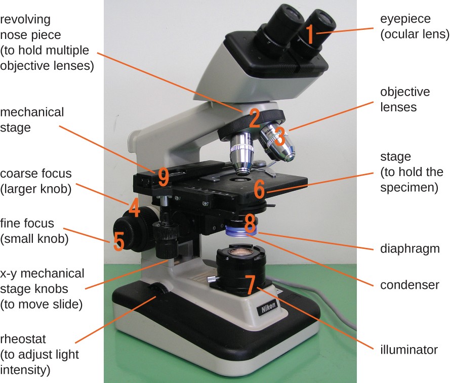
The Parts of a Microscope (Labeled) Printable - TeacherVision The Parts of a Microscope (Labeled) Printable. Download. Add to Favorites. Share. This diagram labels and explains the function of each part of a microscope. Use this printable as a handout or transparency to help prepare students for working with laboratory equipment. Microscope picture label Flashcards | Quizlet Microscope picture label Flashcards | Quizlet Microscope picture label STUDY Flashcards Learn Write Spell Test PLAY Match Gravity Created by kfire Terms in this set (12) Arm What is the part labelled C? Base What is the part labelled D? Body tube What is the part labelled B? Ocular lens What is the part labelled A? Illuminator Explanation and Labelled Images - New York Microscope Company The samples are labeled with fluorophore where they absorb the high-intensity light from the source and emit a lower energy light of longer wavelength. The resulting fluorescent light is then separated from the surrounding radiation with filters, allowing the observer to see only the fluorescing material.
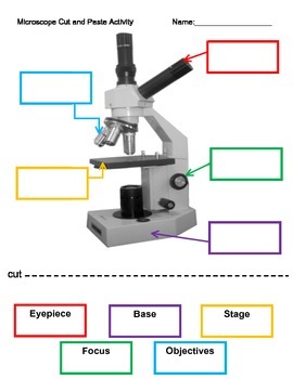
Microscope images with labels. Microscope Label Worksheets & Teaching Resources | TpT Label the Microscope and Magnification Reading PassageIncluded in the resource:A reading passageA student question sheetAn answer keyReading Passage:Included is an informative, introductory passage. The passage has key information around the subject and explains key concepts. With an image of a micr Microscope Labeling - The Biology Corner Students label the parts of the microscope in this photo of a basic laboratory light microscope. Can be used for practice or as a quiz. ... 20. A microscope has an ocular objective of 10x and a high power objective of 50x, what is the microscope's total magnification? _____ PDF Label parts of the Microscope Label parts of the Microscope: . Created Date: 20150715115425Z 18,634 Microscope drawing Images, Stock Photos & Vectors - Shutterstock Find Microscope drawing stock images in HD and millions of other royalty-free stock photos, illustrations and vectors in the Shutterstock collection. Thousands of new, high-quality pictures added every day.
Microscope Parts, Function, & Labeled Diagram - slidingmotion Microscope parts labeled diagram gives us all the information about its parts and their position in the microscope. Microscope Parts Labeled Diagram The principle of the Microscope gives you an exact reason to use it. It works on the 3 principles. Magnification Resolving Power Numerical Aperture. Parts of Microscope Head Base Arm Eyepiece Lens Amazing 27 Things Under The Microscope With Diagrams The tail is transparent and thus is difficult to detect under a low-power microscope. 23. Spirogyra under the microscope. Spirogyra is a green alga found mostly in freshwater in the form of green clumps. Spirogyra is unicellular, but because it clumps together, it can be seen in the pond even with our naked eyes. Microscope, Microscope Parts, Labeled Diagram, and Functions Microscope, Microscope Parts, Labeled Diagram, and Functions What is Microscope? A microscope is a laboratory instrument used to examine objects that are too small to be seen by the naked eye. It is derived from Ancient Greek words and composed of mikrós, "small" and skopeîn,"to look" or "see". Simple Microscope - Diagram (Parts labelled), Principle, Formula and Uses A simple microscope consists of Optical parts Mechanical parts Labeled Diagram of simple microscope parts Optical parts The optical parts of a simple microscope include Lens Mirror Eyepiece Lens A simple microscope uses biconvex lens to magnify the image of a specimen under focus.
Parts of a Simple Microscope - Labeled (with diagrams) image 2: A simple microscope commonly used by students for studying minute objects. image source: imimg.com. picture 3: It is the latest design of a simple microscope - advanced features than the conventional simple microscopes. ... image 5: A modern simple microscope with the different parts labeled. image source: laboratoryinfo.com. The ... 300+ Free Microscope & Laboratory Images - Pixabay Upload 399 Free images of Microscope Related Images: laboratory science bacteria research scientist lab biology chemistry medical Find your perfect microscope image. Free pictures to download and use in your next project. 399 Free images of Microscope / 4‹ › Microscope Types (with labeled diagrams) and Functions This is an advanced microscope that has specific application in viewing, observing and measuring the optical thickness and phase of completely transparent specimens and objects. A tiny interferometer is used and a specimen is placed on beam path of it. This path is split and then rejoined to create two superimposed images of the specimen in focus. Compound Microscope Parts - Labeled Diagram and their Functions The eyepiece (or ocular lens) is the lens part at the top of a microscope that the viewer looks through. The standard eyepiece has a magnification of 10x. You may exchange with an optional eyepiece ranging from 5x - 30x. [In this figure] The structure inside an eyepiece. The current design of the eyepiece is no longer a single convex lens.
Tongue Histology | Histology slides, Human anatomy and physiology, Tissue biology
Microscope Labeling - The Biology Corner The google slides shown below have the same microscope image with the labels for students to copy. I often spend the first day walking students through the steps and having them look at a single slide as we do the steps. Students are often very enthusiastic about using microscopes and will try to start with the high power objective.

Labeling a Compound Microscope Quiz
Parts of the Microscope with Labeling (also Free Printouts) Microscopes are specially created to magnify the image of the subject being studied. This exercise is created to be used in homes and schools. the microscope layout, including the blank and answered versions are available as pdf downloads. Click to Download : Label the Parts of the Microscope (A4) PDF print version.

Best Top Desktop Wallpapers: Electron microscope images
Parts of a microscope with functions and labeled diagram Optical parts of a microscope and their functions The optical parts of the microscope are used to view, magnify, and produce an image from a specimen placed on a slide. These parts include: Eyepiece - also known as the ocular. This is the part used to look through the microscope. Its found at the top of the microscope.

32 Label Of Compound Microscope - Label Design Ideas 2020
Microscope Label Interactive Worksheets & Teaching Resources | TpT 12. $1.89. PDF. Students will complete a timeline of the history of the microscope, label a diagram, and create a pocket foldable with terms and definition cards. The timeline can be completed according to the teacher's directions or like the answer key example. Optional cut & paste images and a QR code are a.

Labels Of The Microscope
Simple Microscope - Parts, Functions, Diagram and Labelling Simple Microscope - Parts, Functions, Diagram and Labelling A microscope is one of the commonly used equipment in a laboratory setting. A microscope is an optical instrument used to magnify an image of a tiny object; objects that are not visible to the human eyes. Table of Contents The common types of microscopes are: What is a Simple microscope?
23 Label And Color The Parts Of Both Microscopes - Labels 2021
Microscope Labeled Pictures, Images and Stock Photos Browse 49 microscope labeled stock photos and images available, or start a new search to explore more stock photos and images. Newest results Fluorescent Imaging immunofluorescence of cancer cells growing... Microscope diagram vector illustration. Labeled zoom instrument... Microscope diagram vector illustration.

MICROBIOLOGY SLIDE SPECIMENS
Microscope Diagram and Functions | Science fair projects ... - Pinterest A Study of the Microscope and its Functions With a Labeled Diagram To better understand the structure and function of a microscope, we need to take a look at the labeled microscope diagrams of the compound and electron microscope. These diagrams clearly explain the functioning of the microscopes along with their respective parts. M mooketsi
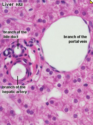
ANAT2241 Liver, Gallbladder, and Pancreas - Embryology
22 Parts Of a Microscope With Their Function And Labeled Diagram Microscope Description A microscope is a laboratory instrument used to examine objects that are too small to be seen by the naked eye. In other words, it enlarges images of small objects. Invented by a Dutch spectacle maker in the late 16th century, light microscopes use lenses and light to magnify images. Generally a microscope ... Read more 22 Parts Of a Microscope With Their Function And ...
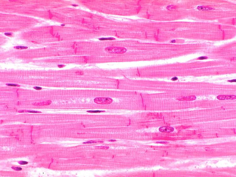
Tissues Flashcards | Easy Notecards
Skin Images Labeled | Virtual Anatomy Lab VAL - ncccval Skin Images Labeled | Virtual Anatomy Lab VAL ... Connective Tissue Images Unlabeled. Microscope. Microscope Images Labeled. Microscope Images Unlabeled. Mitosis. Mitosis Images Labeled. Mitosis Images Unlabeled. Skin. Skin Images Labeled. Skin Images Unlabeled. Skeletal system. Skeletal Images Labeled. Skeletal Images Unlabeled.
Microscope Introduction – “e” Lab - Biology LibreTexts
Labeling the Parts of the Microscope Labeling the Parts of the Microscope This activity has been designed for use in homes and schools. Each microscope layout (both blank and the version with answers) are available as PDF downloads. You can view a more in-depth review of each part of the microscope here. Download the Label the Parts of the Microscope PDF printable version here.

Microscope labeling
Explanation and Labelled Images - New York Microscope Company The samples are labeled with fluorophore where they absorb the high-intensity light from the source and emit a lower energy light of longer wavelength. The resulting fluorescent light is then separated from the surrounding radiation with filters, allowing the observer to see only the fluorescing material.

31 Label The Indicated Parts Of The Microscope - Labels For Your Ideas
Microscope picture label Flashcards | Quizlet Microscope picture label Flashcards | Quizlet Microscope picture label STUDY Flashcards Learn Write Spell Test PLAY Match Gravity Created by kfire Terms in this set (12) Arm What is the part labelled C? Base What is the part labelled D? Body tube What is the part labelled B? Ocular lens What is the part labelled A? Illuminator
32 Picture Of Microscope With Label - Labels For You
The Parts of a Microscope (Labeled) Printable - TeacherVision The Parts of a Microscope (Labeled) Printable. Download. Add to Favorites. Share. This diagram labels and explains the function of each part of a microscope. Use this printable as a handout or transparency to help prepare students for working with laboratory equipment.
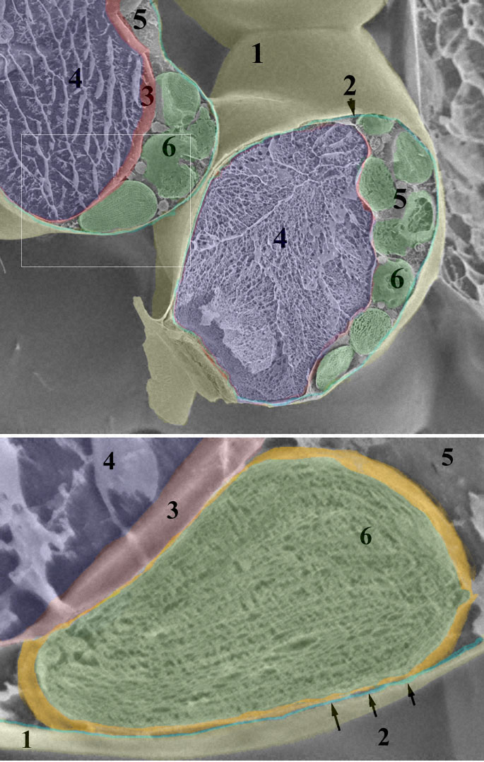
Leaf chloroplast

Microscope labeling
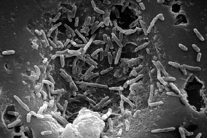
Mycobacterium leprae* - microbewiki

Microscope Clip Art at Clker.com - vector clip art online, royalty free & public domain
Post a Comment for "44 microscope images with labels"