45 eye diagram with labels and functions
Control Unit Installation and Operation Guide Please Read between any Eye QS control unit and any other power supply, including another GRAFIK Eye QS control unit. Refer to the QS Link Power Draw Units specification submittal (Lutron P/N 369405) for more information concerning PDUs. 1234 12 ABC 123456LN Example: Emergency lighting interface (maximum 1) Note: The GRAFIK Eye QS control unit Microscope Types (with labeled diagrams) and Functions Simple microscope labeled diagram Simple microscope functions It is used in industrial applications like: Watchmakers to assemble watches Cloth industry to count the number of threads or fibers in a cloth Jewelers to examine the finer parts of jewelry Miniature artists to examine and build their work Also used to inspect finer details on products
plotly.com › python › parallel-categories-diagramParallel categories diagram in Python - Plotly Basic Parallel Categories Diagram with graph_objects¶ This example illustrates the hair color, eye color, and sex of a sample of 8 people. The dimension labels can be dragged horizontally to reorder the dimensions and the category rectangles can be dragged vertically to reorder the categories within a dimension.

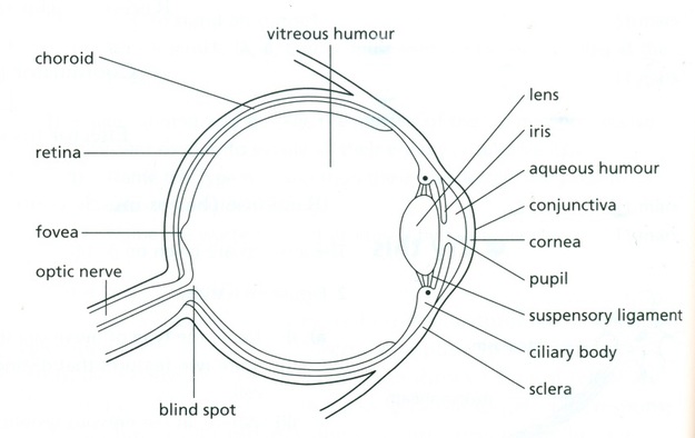
Eye diagram with labels and functions
Eye anatomy: Muscles, arteries, nerves and lacrimal gland - Kenhub Six extraocular muscles move the eye: superior rectus, inferior rectus, medial rectus, lateral rectus, superior oblique and inferior oblique muscles; and one other, levator palpebrae superioris, opens the eyelid. Don't understand how all these muscles work? You can find out everything about them in the following learning materials. Consumer Updates | FDA - U.S. Food and Drug Administration 28.07.2022 · The site is secure. The https:// ensures that you are connecting to the official website and that any information you provide is encrypted and transmitted securely. The Anatomy and Function of a Cat's Eye - PetHelpful The mysterious fluorescent shine of a cat's eyes, when caught in a beam of light, is caused by a structure called the tapetum lucidum, located directly behind the retina. The cat's tapetum works like a mirror, allowing lights to bounce off of it and increase visual sensitivity. It works similarly to those reflective lane markings on the highway.
Eye diagram with labels and functions. Parts of Human Eye and Their Functions | MD-Health.com The different parts of the eye allow the body to take in light and perceive objects around us in the proper color, detail and depth. This allows people to make more informed decisions about their environment. If a portion of the eye becomes damaged, you may not be able to see effectively, or lose your vision all together. What are the parts ? Important Question for Class 10 Science Human Eye and Colourful World Important Questions of Light Reflection and Refraction Class 10 Science Chapter 11. Question 1. State one function of iris in human eye. (AI 2012) Answer: Irish adark muscular diaphragm that controls the size of the pupil. Question 2. State one function of the crystalline lens in the human eye. › labelling_interactives › 6Label the microscope — Science Learning Hub Jun 08, 2018 · Labels. Description. eye piece lens. The lens you look through – normally 10x or 15x magnification. coarse focus adjustment. Moves the lens up or down and adjusts focus. fine focus adjustment. Moves the lens in order to make very small adjustments to gain better focus. base. The bottom of the microscope used for stability. high-power objective Create a Briliant Process Flow Diagram with Canva There are lots of ways to use color in a process flow diagram. You could have all the arrows in one part of the process the same color to make it clear they relate to that process. For example, you could use colors like blue and green to represent a cooling process or red and yellow to represent something being heated. To recolor any element or text in your design, select it, then …
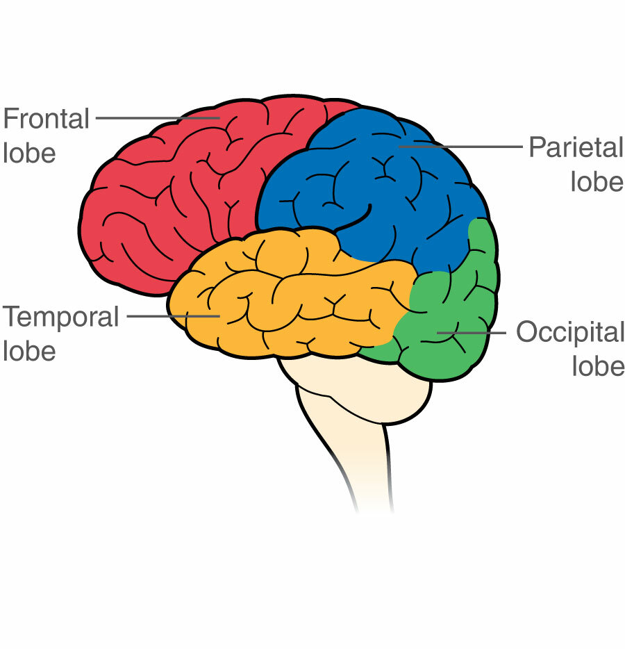
Neuroanatomy, Visual Pathway - StatPearls - NCBI Bookshelf Visual stimuli from our surroundings are processed by an intricate system of interconnecting neurons, which begins with the optic nerve in the eye up to the visual processing center in our forebrain called the visual cortex. All the information travels in the form of nerve impulses that are triggered by photosensitive chemical reactions occurring in the retina. Several separate and parallel ... What are the 12 cranial nerves? Functions and diagram - Medical News Today The functions of the cranial nerves are sensory, motor, or both. Sensory cranial nerves help a person see, smell, and hear. Conversely, motor cranial nerves help control muscle movements in the... Human eye - Wikipedia The human eye is a sensory organ, part of the sensory nervous system, that reacts to visible light and allows us to use visual information for various purposes including seeing things, keeping our balance, and maintaining circadian rhythm.. The eye can be considered as a living optical device.It is approximately spherical in shape, with its outer layers, such as the outermost, white … Microscope Parts, Function, & Labeled Diagram - slidingmotion Microscope parts labeled diagram gives us all the information about its parts and their position in the microscope. Microscope Parts Labeled Diagram The principle of the Microscope gives you an exact reason to use it. It works on the 3 principles. Magnification Resolving Power Numerical Aperture. Parts of Microscope Head Base Arm Eyepiece Lens
Mesencephalon | Structure, Position, Function & Facts The mesencephalon of the midbrain is part of the brain located in the most rostral part of the Truncus encephali or the brain stem, between the hindbrain and the forebrain ().Mesencephalon connects these two parts of the brain. At the same time, this is the most superior brain region located in the brainstem. Its key peculiarity is numerous nerve tracts that play the key role in connecting the ... Generate eye diagram - MATLAB eyediagram - MathWorks eyediagram(x,n) generates an eye diagram for signal x, plotting n samples in each trace.The labels on the horizontal axis of the diagram range between –1/2 and 1/2. The function assumes that the first value of the signal and every nth value thereafter, occur at integer times. Eye Anatomy | Blood supply - Orbit - Extraocular muscles - Geeky Medics There are three fluid-filled areas within the eye: the anterior chamber, posterior chamber and vitreous body. The anterior and posterior chambers contain aqueous humour, which is constantly produced and drained from the eye. Impaired drainage of aqueous humour can result in glaucoma The vitreous body contains vitreous humour The extraocular muscles Oscilloscope Feature and Options Table - Siglent Here is a table of features and options of our most powerful oscilloscopes to help decide what is best for you. (2) - Requires SIGLENT SAG hardware and activation license. The SDS2X Plus series has an internal function generator but still requires activation. For convenience, click a link below to jump to the product of interest:
Human Eye Diagram Labeled - Health, Medicine and Anatomy Reference Pictures | School | Pinterest ...
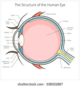
Anatomy and Structure of the Human Eye (With Diagrams) In the diagram, the artery is shown in red, and the vein is in blue. Tear Duct This is a small tube running from the eye to the nasal cavity. Tears drain from the eyes into the nose through the tear duct. This is why teary eyes are usually accompanied by a runny nose. Schematic animation of the human eyeball
Labeled Picture Of The Eye - ClipArt Best
› TechnicalDocumentLibrary › GrafikControl Unit Installation and Operation Guide Please Read between any Eye QS control unit and any other power supply, including another GRAFIK Eye QS control unit. Refer to the QS Link Power Draw Units specification submittal (Lutron P/N 369405) for more information concerning PDUs. 1234 12 ABC 123456LN Example: Emergency lighting interface (maximum 1) Note: The GRAFIK Eye QS control unit
Document Moved
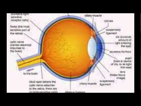
Anatomy of the Eye | BrightFocus Foundation Glossary of Terms. Anterior chamber: The region of the eye between the cornea and the lens that contains aqueous humor. Aqueous humor: The fluid produced in the eye. Bruch's membrane: Located in the retina between the choroid and the retinal pigmented epithelium (RPE) layer; provides support to the retina and functions as the 'basement' membrane of the RPE layer.

Can anyone pls help me with an eye (fully labelled)diagram...it's given wrong in our book.Kindly ...
Conjunctiva: Anatomy, Function, and Treatment - Verywell Health Conjunctivitis, also known as pink eye , is inflammation or infection of the conjunctiva. 1 Some forms (bacterial, viral) are highly contagious. Other forms may be triggered by an allergy or exposure to harsh chemicals. Symptoms can be persistent and include redness, itching, tearing, discharge, and more. Verywell / Emily Roberts Is It Pink Eye?

Learning English in a new way: diciembre 2013
Quiz: Label The Parts Of The Eye - ProProfs Quiz How much did you get to understand about the human eye? Take up this quiz and find out! Questions and Answers. 1. A is pointing to what part of the eye? A. Cornea. B. Optic Nerve.

Diagram Of An Eye And Its Functions - Diagram Media
Liver Diagram with Detailed Illustrations and Clear Labels - BYJUS Liver – Anatomy, Functions, And Liver Diseases; Also Read: 6 Facts Everyone Should Know About The Liver; Fatty Liver Symptoms – Explore The Signs, Indications And Causes; 12 Alarming Symptoms of Liver Problems You Shouldn’t Ignore; A Brief Account Of Hepatic Portal System And Its Significance; Human Body – Anatomy and Physiology of ...

Module 1: Labeled Diagram of the Eye | Eye health | Pinterest
Microscope, Microscope Parts, Labeled Diagram, and Functions Microscope, Microscope Parts, Labeled Diagram, and Functions What is Microscope? A microscope is a laboratory instrument used to examine objects that are too small to be seen by the naked eye. It is derived from Ancient Greek words and composed of mikrós, "small" and skopeîn,"to look" or "see".
Structure And Function Of The Eye - YouTube
Parts Of The Eye Quiz Questions And Answers - ProProfs Quiz Questions and Answers 1. What is the best definition of the retina? A. An outermost part of the eye that sees things. B. The innermost part of the eye that absorbs light and changes it into electrical signals. C. The light switch to the brain. D. The clear, tough tissue covering the front of the eye. 2.
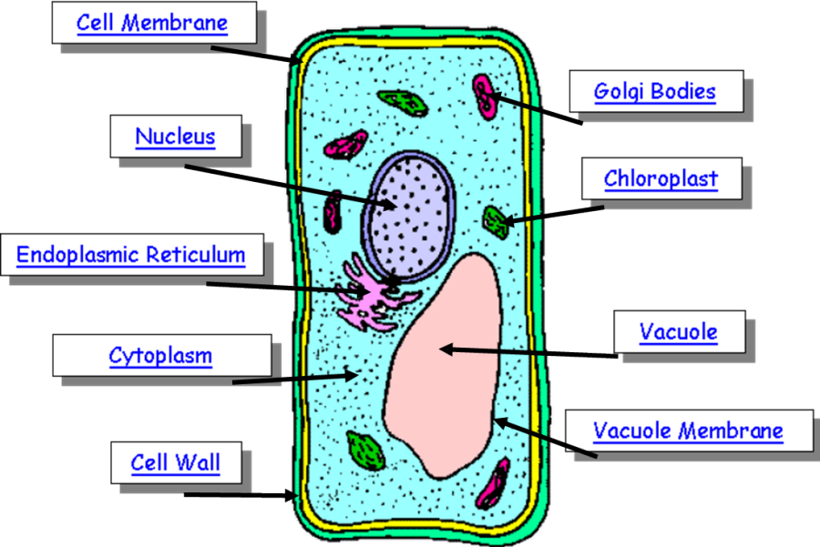
Plant Cell Drawing With Labels at PaintingValley.com | Explore collection of Plant Cell Drawing ...
Excel Gauge Chart Template - Free Download - How to Create Move the labels to the appropriate places above the gauge chart. Change the chart title. Bonus Step for the Tenacious: Add a text box with your actual data value. Here is a quick and dirty tip on making the speedometer chart more informative as well as pleasing to the eye. Let’s add a text box that will display the actual value of the pointer.

Functions and Anatomy of the Eye by Health EDventure | TpT
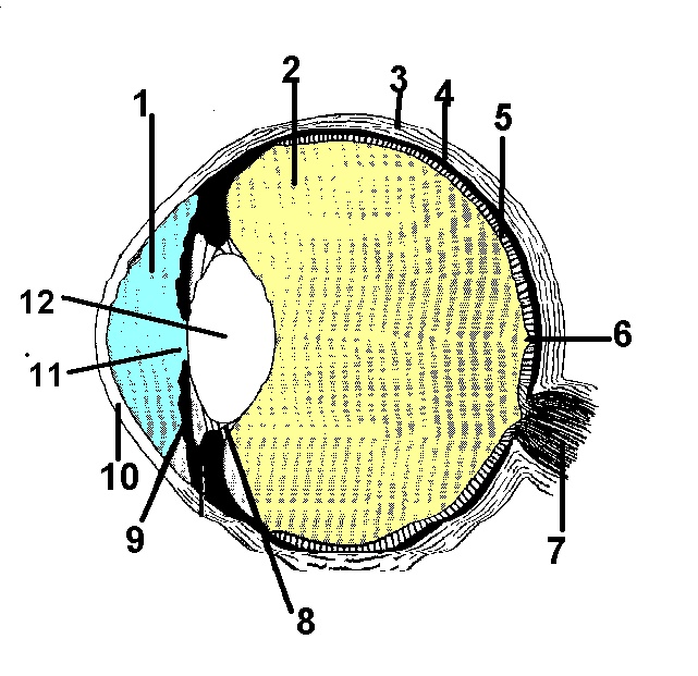
Parts Of The Eye Labeled Diagram Model And Their Function Parts of the eye-labeled diagram model are divided into three groups: the external outer layer, the middle layer, and the inner back layer. The outer layer is responsible for protecting the eye from environmental toxins and debris. The middle layer includes cells that allow light to enter and travel through the back layer to the retina.
The eye, rods and cones - Biology Notes for IGCSE 2014
Human Eye - Light - Class Notes Function of Iris and Pupil The iris automatically adjusts the size of pupil according to the intensity of light received by the eye from the surroundings. (1) If the amount of light around us is very high (as during the day light), then iris contracts the pupil (makes the pupil small) and hence reduces the amount of light entering the eye.

Draw a labelled diagram of the human eye. Label the following parts on this diagram:
Anatomy of the eye: Quizzes and diagrams | Kenhub One of our favorite ways to get to grips with all of the parts of the eye is by utilizing labeled diagrams. On a diagram of the eye, we can see all of the relevant structures together on one image. This helps us to understand how each one is situated and related to the other. Labeled diagram of the eye
Easy Human Eye Diagram With Labels - Diagram Media
The Lens: Anatomy, Function, and Treatment - Verywell Health Function Associated Conditions Tests The lens is a curved structure in the eye that that bends light and focuses it for the retina to help you see images clearly. The crystalline lens, a clear disk behind the iris, is flexible and changes shape to help you see objects at varying distances. As you age, the lens may become weaker or damaged.

Main veins of the leg stock vector. Illustration of iliac - 121325793
Label the microscope — Science Learning Hub 08.06.2018 · All microscopes share features in common. In this interactive, you can label the different parts of a microscope. Use this with the Microscope parts activity to help students identify and label the main parts of a microscope and then describe their functions.. Drag and drop the text labels onto the microscope diagram. If you want to redo an answer, click on the box and …
Diagram of the Eye | ClipArt ETC
20/20 Vision - Activity - TeachEngineering Require the diagram to include a light source, an object to be seen, and the eye viewing the object. In the diagram, have them label the cornea, iris and pupil, lens, and retina. Additionally, challenge students to add a second lens (glasses) to the diagram and describe the effect on vision of the added lens. Troubleshooting Tips
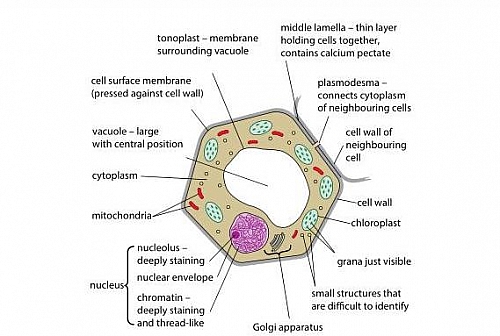
Plant cell Structure: Plant cell parts, Organelles and their functions and Diagram - Jotscroll
Eye Anatomy and Physiology a Complete Detail - Study Read Eye Anatomy and Physiology Eyes are spheroid shape organs fitted into the two orbitals of the skull. There are three major parts in each eye like The sclera (fibrous layer) Choroid layer Retina Eyes diagram showing the entire structure The sclera It makes up the outermost part of eye anatomy.
Post a Comment for "45 eye diagram with labels and functions"