41 human heart and labels
› coronavirus › 2019-ncovMyocarditis and Pericarditis After mRNA COVID-19 Vaccination Sep 27, 2022 · Myocarditis is inflammation of the heart muscle. Pericarditis is inflammation of the outer lining of the heart. In both cases, the body’s immune system causes inflammation in response to an infection or some other trigger. Learn more about myocarditis and pericarditis. Both myocarditis and pericarditis have the following symptoms: Chest pain › heart › picture-of-the-heartHuman Heart (Anatomy): Diagram, Function, Chambers, Location ... Heart Treatments. Exercise: Regular exercise is important for heart health and most heart conditions.Talk to your doctor before starting an exercise program if you have heart problems. Angioplasty ...
commons.wikimedia.org › wiki › File:Diagram_of_theFile : Diagram of the human heart (cropped).svg - Wikimedia Add Inferior vena cava and pericardium labels: 18:08, 14 August 2018: 656 × 631 (209 KB) Jmarchn (talk | contribs) Add pericardium. Add papillary muscles and chordae tendinae. Add cardiac skeleton. ... Diagram of the human heart, created by Wapcaplet in Sodipodi. Cropped by ~~~ to remove white space (this cropping is not the same as Wapcaplet ...
Human heart and labels
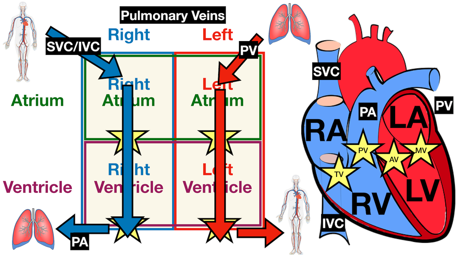
Human Heart - Diagram and Anatomy of the Heart - Innerbody The heart contains 4 chambers: the right atrium, left atrium, right ventricle, and left ventricle. The atria are smaller than the ventricles and have thinner, less muscular walls than the ventricles. The atria act as receiving chambers for blood, so they are connected to the veins that carry blood to the heart. heart | Structure, Function, Diagram, Anatomy, & Facts heart, organ that serves as a pump to circulate the blood. It may be a straight tube, as in spiders and annelid worms, or a somewhat more elaborate structure with one or more receiving chambers (atria) and a main pumping chamber (ventricle), as in mollusks. In fishes the heart is a folded tube, with three or four enlarged areas that correspond to the chambers in the mammalian heart. The Human Heart Labeling Worksheet - Twinkl The human heart is a muscle made up of four chambers, these are: Two upper chambers - the left atrium and right atrium Two lower chambers - the left and right ventricles. It's also made up of four valves - these are known as the tricuspid, pulmonary, mitral and aortic valves.
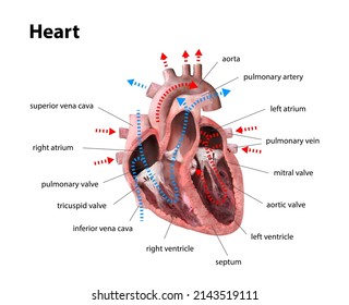
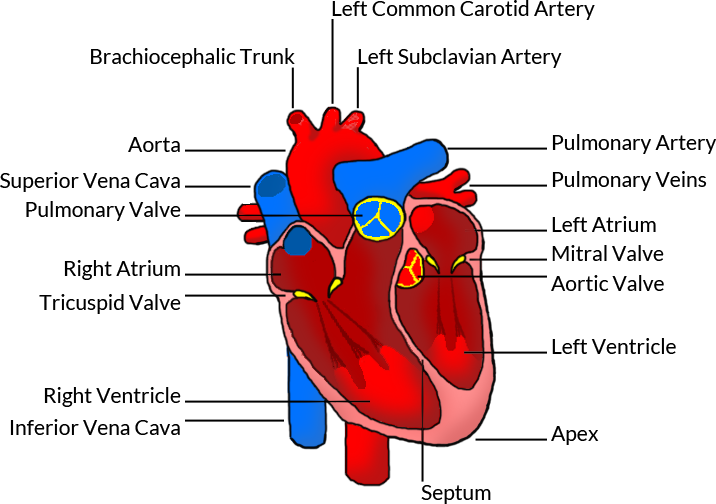
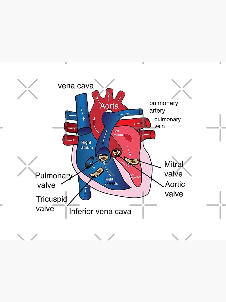
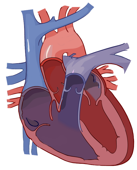
Human heart and labels. Human Heart Diagram Pictures, Images and Stock Photos Browse 4,129 human heart diagram stock photos and images available, or search for heart illustration or pulmonary artery to find more great stock photos and pictures. heart anatomy. Part of the human heart. Human heart cross section, cardiovascular system diagram isolated on white. Human Heart Diagram - Human Body Pictures - Science for Kids Photo description: This is an excellent human heart diagram which uses different colors to show different parts and also labels a number of important heart component such as the aorta, pulmonary artery, pulmonary vein, left atrium, right atrium, left ventricle, right ventricle, inferior vena cava and superior vena cava among others. The Anatomy of the Heart, Its Structures, and Functions - ThoughtCo The heart is the organ that helps supply blood and oxygen to all parts of the body. It is divided by a partition (or septum) into two halves. The halves are, in turn, divided into four chambers. The heart is situated within the chest cavity and surrounded by a fluid-filled sac called the pericardium. This amazing muscle produces electrical ... Label the heart — Science Learning Hub In this interactive, you can label parts of the human heart. Drag and drop the text labels ...
Parts Of The Human Heart | Science Trends The parts of the human heart can be broken down into four chambers, muscular walls, vessels, and a conductive system. The two upper chambers are called the atria, with lower parts called ventricles. These all work together to make up the vital function of your heart. Everybody knows that the human heart is the essential organ in our bodies. Labelling the heart — Science Learning Hub Labelling the heart. The heart is a muscular organ that pumps blood through the blood vessels of the circulatory system. Blood transports oxygen and nutrients to the body. It is also involved in the removal of metabolic wastes. In this activity, students use online and paper resources to identify and label the main parts of the heart. Heart Labeling Quiz: How Much You Know About Heart Labeling? Create your own Quiz Here is a Heart labeling quiz for you. The human heart is a vital organ for every human. The more healthy your heart is, the longer the chances you have of surviving, so you better take care of it. Take the following quiz to know how much you know about your heart. Questions and Answers 1. What is #1? 2. What is #2? 3. Human Heart Labeling Teaching Resources | Teachers Pay Teachers Perfect for middle school or high school biology.Includes:- 1 diagram of the human heart & its vessels, unlabeled;- 1 diagram labeled 1-18 with lines for student answers;- 1 diagram labeled A-R with parts of the heart & its vessels identified for matching purposes;- 1 diagram labeled A-R;- 1 diagram labeled with
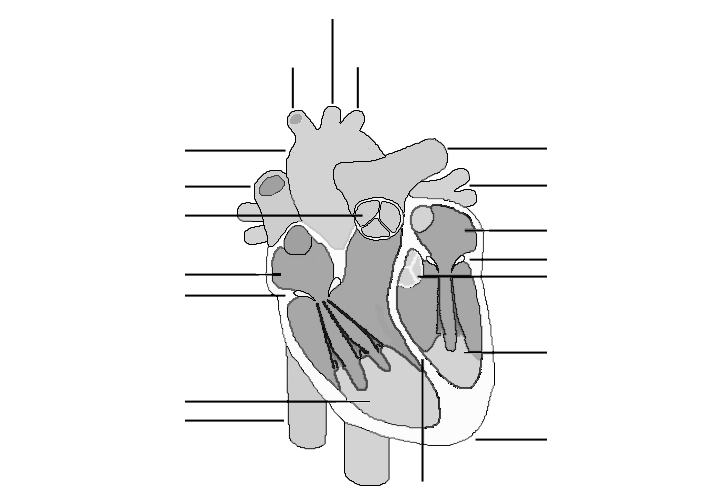
Diagram of the human heart royalty-free images - Shutterstock Find Diagram of the human heart stock images in HD and millions of other royalty-free stock photos, illustrations and vectors in the Shutterstock collection. Thousands of new, high-quality pictures added every day. Unlabelled Heart Diagram - Primary Biology Resources This fab Unlabelled Heart Diagram worksheet is a great way to help your primary kids with their learning of the basic human anatomy. On the downloadable PDF is a handy heart diagram without labels, so your students can label it themselves and familiarise themselves with the different parts of the human heart. Heart Diagram with Labels and Detailed Explanation - BYJUS Well-Labelled Diagram of Heart. The heart is made up of four chambers: The upper two chambers of the heart are called auricles. The lower two chambers of the heart are called ventricles. The heart wall is made up of three layers: The outer layer of the heart wall is called epicardium. The middle layer of the heart wall is called myocardium. The inner layer of the heart wall is called endocardium. Label the Human Heart | eCampusOntario H5P Studio Drag and drop the labels to identify the different parts of the human heart. Available for adopting/adapting. ... Medicine & Nursing. Administrative Title Label the Human Heart. H5P ID 191. Last Update 2 years ago. Created 04 Mar 2020. License Error: Failed to load License Attribution. Sarah Wendorf. Instructional Designer, Cambrian College ...
652 Human Heart 3d With Label Images, Stock Photos & Vectors ...
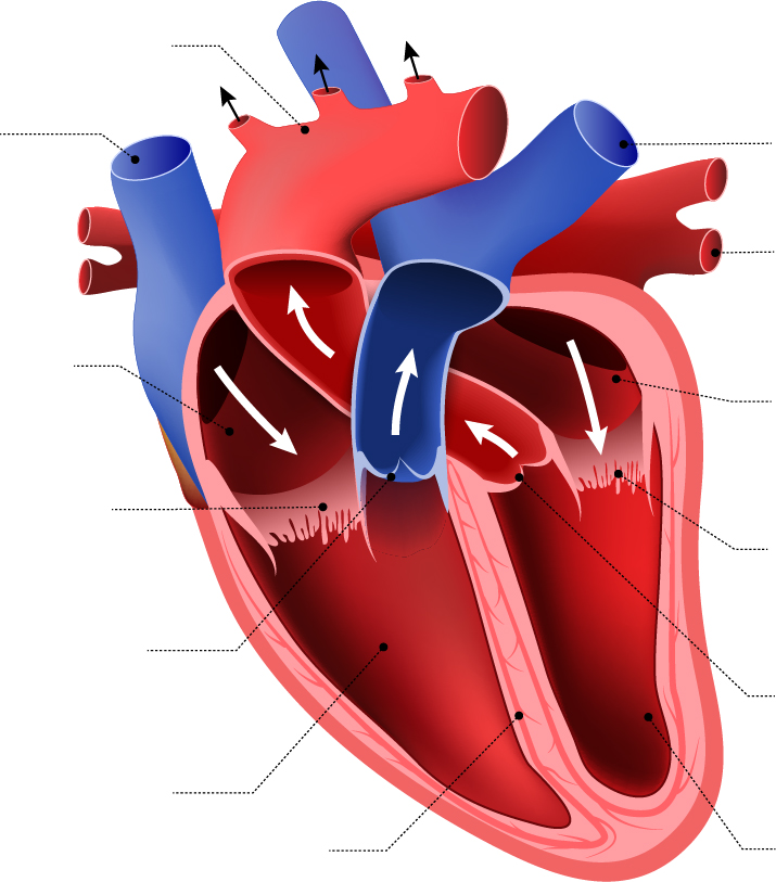
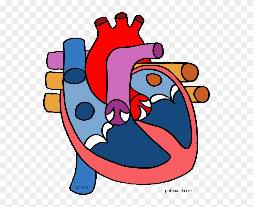
How to Draw a Human Heart: An Easy Step-By-Step Guide - wikiHow Generally, hearts on anatomical or medical diagrams are sectioned into 2 colors: red and blue. The red represents blood going into the heart, whereas the blue represents blood leaving the heart. [8] Split the heart in half and color the left section, superior vena cava, and pulmonary artery blue, and then color the right section and aorta red. [9]

How to draw Human heart | Human heart | Quickly | Well labelled diagram | step by step, NCERT |
Human Heart Diagram Without Labels - Labelling Worksheet - Twinkl The human heart is a muscle made up of four chambers, these are: Two upper chambers - the left atrium and right atrium Two lower chambers - the left and right ventricles. It's also made up of four valves - these are known as the tricuspid, pulmonary, mitral and aortic valves.
a) Draw a sectional view of the human heart and label on it ...
Human Heart with Labels on White Background stock photo ... iStock Human Heart With Labels On White Background Stock Photo - Download Image Now - Right Atrium, Diagram, Human Heart Download this Human Heart With Labels On White Background photo now. And search more of iStock's library of royalty-free stock images that features Right Atrium photos available for quick and easy download.

Describe the structure of the heart of human with the help of ...
Human Heart Diagram Labeled | Science Trends Human Heart Diagram Labeled Anatomy Of The Heart. The human heart usually weighs somewhere between 10 to 12 ounces in men and between 8 to 10 ounces... Pumping Blood Through The Body. The circulatory system surrounding the heart. ... The heart's primary function is to... List Of Heart Structures. ...
The Human Heart
human heart and labels human heart and labels Medal Clipart Medal Valor - Purple Heart Military Award , Free. 9 Images about Medal Clipart Medal Valor - Purple Heart Military Award , Free : Human Anatomy Lab: Heart Models, Human Heart Pictures with Labels Best Of File Diagram Of the Human and also Typical Thoracic vertebra - Heart shaped body | Thoracic vertebrae.
Heart Diagram Labelling Activity - Science - Twinkl QR
greenroads.com › collections › cbd-edibles-gummiesBuy CBD Gummies & CBD Edibles Online | Green Roads Buy the CBD gummies and edibles you need at Green Roads! Enjoy lab-tested, pharmacist formulated CBD gummy bears, fruit bites, & sleep gummies with melatonin.

Label the Heart Diagram | Quizlet
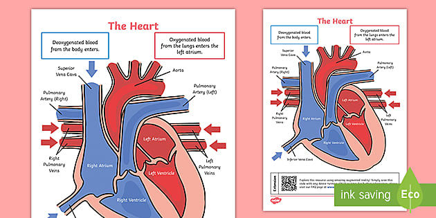
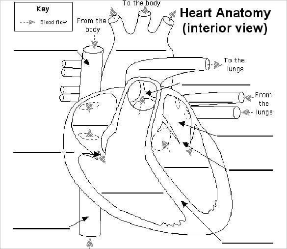
The Human Heart Cardiovascular System Labeling Worksheet - Twinkl This handy heart worksheet gives your children the opportunity to show how much they've learned about this topic. Using the blank heart diagram students are asked to label the aorta, superior vena cava, pulmonary arteries, pulmonary veins, atrium, ventricles, and aortic valves. This simple human heart diagram could be used as both a starter or plenary in order to assess students ...
File:Diagram of the human heart (cropped)-it.png - Wikimedia ...
26,219 Human heart label Images, Stock Photos & Vectors - Shutterstock Find Human heart label stock images in HD and millions of other royalty-free stock photos, illustrations and vectors in the Shutterstock collection. Thousands of new, high-quality pictures added every day.

Sketch and Label Ventral View of Human Heart. - Circulatory ...
A Labeled Diagram of the Human Heart You Really Need to See The human heart, comprises four chambers: right atrium, left atrium, right ventricle and left ventricle. The two upper chambers are called the left and the right atria, and the two lower chambers are known as the left and the right ventricles. The two atria and ventricles are separated from each other by a muscle wall called 'septum'.

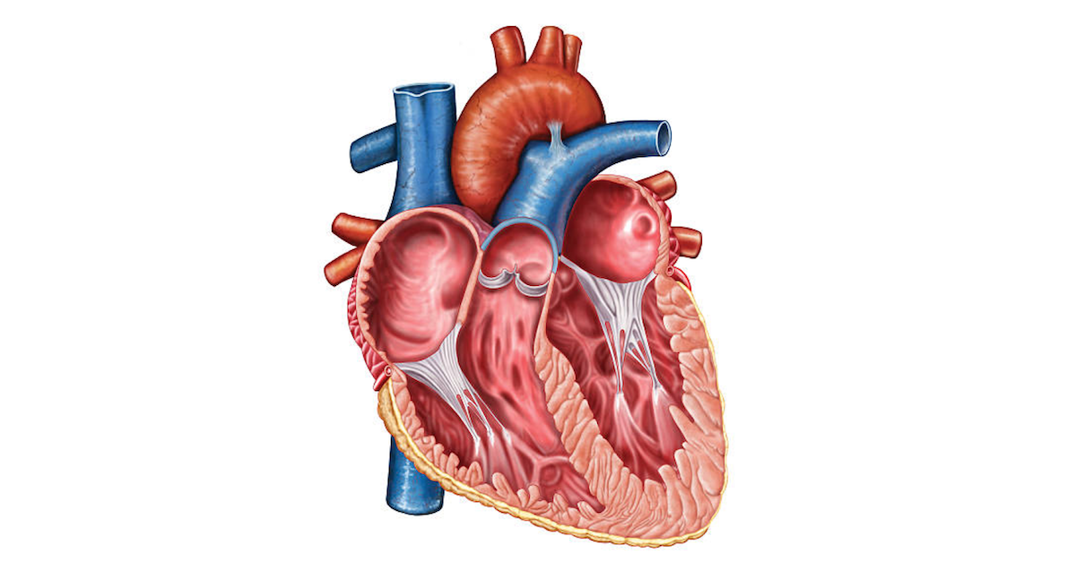
Anatomy of a Human Heart
abcnews.go.com › healthHealth News | Latest Medical, Nutrition, Fitness News - ABC ... Sep 25, 2022 · Get the latest health news, diet & fitness information, medical research, health care trends and health issues that affect you and your family on ABCNews.com

Simple heart diagram | Simple heart diagram labeled | Human ...
heart human label 2.3.1 draw and label a diagram of the ultrastructure of a liver cell as. 9 pics about 2.3.1 draw and label a diagram of the ultrastructure of a liver cell as : human heart pictures with labels best of file diagram of the human, parts of heart diagram stock illustration - download image now - istock and also parts of heart diagram stock …
Heart Anatomy: Labeled Diagram, Structures, Blood Flow ...
human heart and labels human heart and labels Typical Thoracic vertebra - Heart shaped body | Thoracic vertebrae. 9 Pics about Typical Thoracic vertebra - Heart shaped body | Thoracic vertebrae : Human Anatomy Lab: Heart Models, Human Heart Pictures with Labels Best Of File Diagram Of the Human and also Medal Clipart Medal Valor - Purple Heart Military Award , Free.

Human Heart Diagram Labeled | Science Trends
› laws-regs › regulations1910.1030 - Bloodborne pathogens. | Occupational Safety and ... Warning labels shall be affixed to containers of regulated waste, refrigerators and freezers containing blood or other potentially infectious material; and other containers used to store, transport or ship blood or other potentially infectious materials, except as provided in paragraph (g)(1)(i)(E), (F) and (G).

Heart Structure | BioNinja
Diagram of Human Heart and Blood Circulation in It Exterior of the Human Heart. A heart diagram labeled will provide plenty of information about the structure of your heart, including the wall of your heart. The wall of the heart has three different layers, such as the Myocardium, the Epicardium, and the Endocardium. Here's more about these three layers. Epicardium

draw a diagram of the front view oh human heart and label and ...
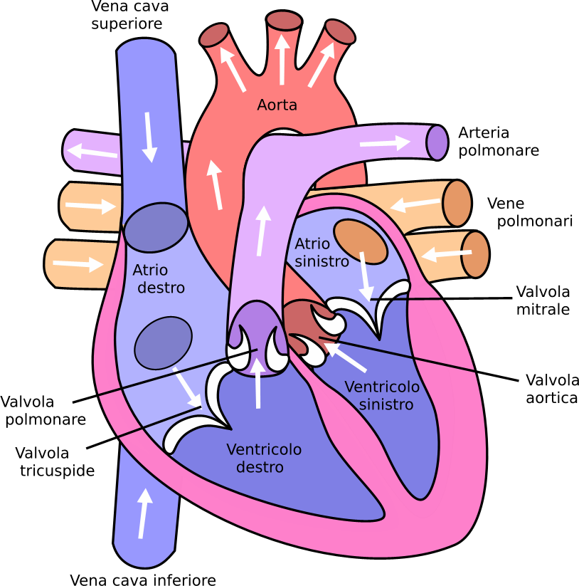
byjus.com › biology › human-heartHuman Heart - Anatomy, Functions and Facts about Heart - BYJUS The human heart is divided into four chambers, namely two ventricles and two atria. The ventricles are the chambers that pump blood and atrium are the chambers that receive the blood. Among which, the right atrium and ventricle make up the "right portion of the heart", and the left atrium and ventricle make up the "left portion of the heart." 5.
The Human Heart
The Human Heart Labeling Worksheet - Twinkl The human heart is a muscle made up of four chambers, these are: Two upper chambers - the left atrium and right atrium Two lower chambers - the left and right ventricles. It's also made up of four valves - these are known as the tricuspid, pulmonary, mitral and aortic valves.
Label the Human Heart | eCampusOntario H5P Studio
heart | Structure, Function, Diagram, Anatomy, & Facts heart, organ that serves as a pump to circulate the blood. It may be a straight tube, as in spiders and annelid worms, or a somewhat more elaborate structure with one or more receiving chambers (atria) and a main pumping chamber (ventricle), as in mollusks. In fishes the heart is a folded tube, with three or four enlarged areas that correspond to the chambers in the mammalian heart.
Heart Anatomy: Labeled Diagram, Structures, Blood Flow ...
Human Heart - Diagram and Anatomy of the Heart - Innerbody The heart contains 4 chambers: the right atrium, left atrium, right ventricle, and left ventricle. The atria are smaller than the ventricles and have thinner, less muscular walls than the ventricles. The atria act as receiving chambers for blood, so they are connected to the veins that carry blood to the heart.

Sketch Human Heart Vector & Photo (Free Trial) | Bigstock
poster of human heart anatomy with hand written labels of the ...
poster of human heart anatomy with hand written labels of the ...
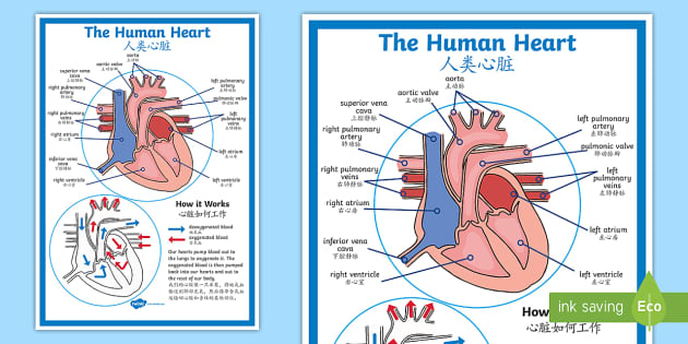
FREE! - The Human Heart Diagram Display Poster - English ...

Human Heart With Labels iPhone 6 Case by Hank Grebe | Pixels
Human Heart Clipart - Heart Diagram No Labels - Free ...

Q1 Given alongside is a diagram of human heart showing its ...
(230).jpg)
Heart Labeling Quiz: How Much You Know About Heart Labeling ...

Heart Anatomy: Labeled Diagram, Structures, Blood Flow ...
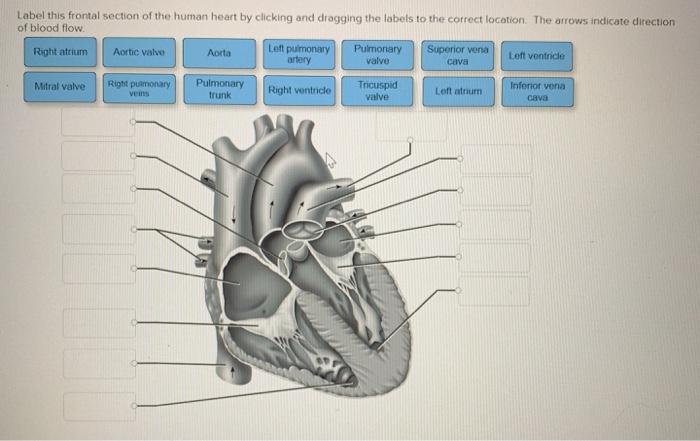
Solved Label this frontal section of the human heart by ...

Human Heart: Label the diagram 1 worksheet
13+ Heart Diagram Templates – Sample, Example, Format ...
652 Human Heart 3d With Label Images, Stock Photos & Vectors ...

Science worksheets: Label parts of a human heart

Heart Anatomy: Labeled Diagram, Structures, Blood Flow ...
Human Heart - Label the Diagram Anatomy Clip Art Set Commercial Use

Human Heart Diagram Without Labels | Human heart diagram ...

Label parts of the heart worksheet

Human Heart Anatomy Infographic Diagram Stock Vector ...
Label the heart — Science Learning Hub
human heart without label - Clip Art Library
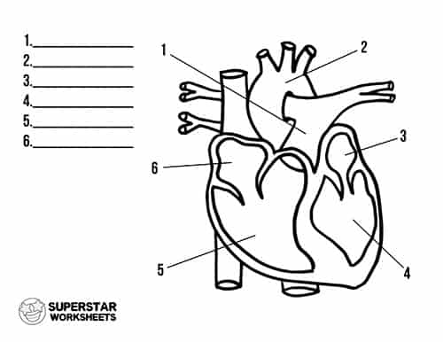
Heart Worksheets - Superstar Worksheets
13+ Heart Diagram Templates – Sample, Example, Format ...
Post a Comment for "41 human heart and labels"